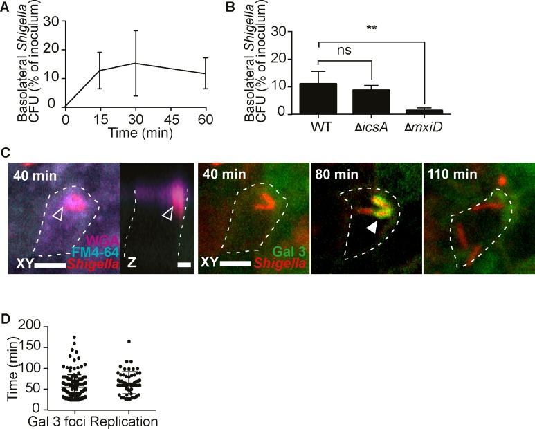
Fig 2. S. flexneri follows a bimodal itinerary within M cells.
(A) S. flexneri translocates rapidly to basolateral compartments (t15, 30 n = 5, t60 n = 6). (B) S. flexneri transcytosis is dependent on bacterial invasion and independent of ABM at 1h p.i.,(WT n = 5, ΔicsA and ΔmxiD n = 4, *P <0.01). (C) S. flexneri induces apical membrane ruffling at 40 min p.i. in a galectin 3-eGFP reporter M cell (delineated by a dashed line), visible in the FM4-64 and WGA channels (empty arrows), before rupturing its vacuole at 80 min p.i. visible by the formation of a galectin 3 cage around the bacteria, and cytosolic replication at 110 min pi, visualized by time-lapse microscopy. Representative time-lapse (n = 3), at different Z-planes (based upon the best view of the different infection events), in the same XY field. Scale bar, 5 μm. See also video S1. (D) Quantification of the onset of S. flexneri vacuolar rupture and replication within M cells imaged live (Gal 3 foci n = 139, replication n = 51, data from three independent experiments).

