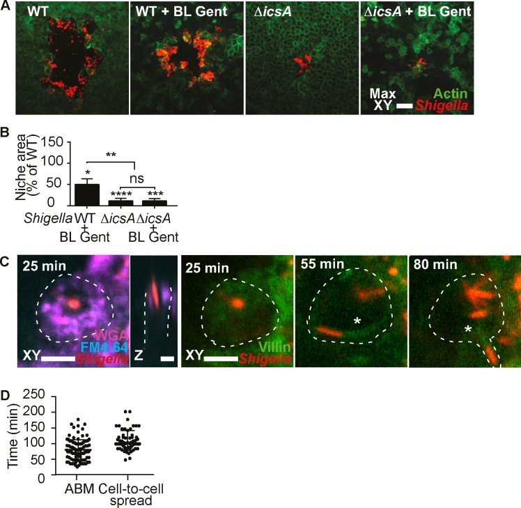
Fig 3. S. flexneri spreads directly from the M cell cytosol to neighboring enterocytes via IcsA.
(A) S. flexneri infection plaques formed in the indicated conditions and strains, visualized by confocal microscopy at 21h p.i.. WT = wild-type, gent = basolateral gentamicin. Scale bar, 50 μm. (B) Quantification of infection plaque areas for the indicated conditions, normalized to WT. The statistical significance of differences between conditions and WT were assessed by one sample t tests, and differences between conditions were assessed by one-way ANOVA and multiple comparisons (WT n = 61, WT + BL Gent n = 40, ΔicsA n = 141, ΔicsA + BL Gent n = 121, WT and ΔicsA data from five independent experiments, + BL Gent data from three independent experiments, (*p < 0.05, **p < 0.01, ****p < 0.0001) (C) S. flexneri induces apical membrane ruffling at 25 min p.i. in a villin-eYFP reporter M cell (delineated by a dashed line), before developing ABM at 55 min p.i., visible by the villin-positive comet tail (asterisk), leading to the development of a membrane protrusion visible by FM4-64 labeling and direct spread at 80 min p.i. into a neighboring cell. Scale bar, 5 μm. See also video S2. (D) Quantification of the onset of S. flexneri ABM and cell-to-cell spread from M cells (ABM n = 93, Cell-to-cell spread n = 46, data from three independent experiments).

