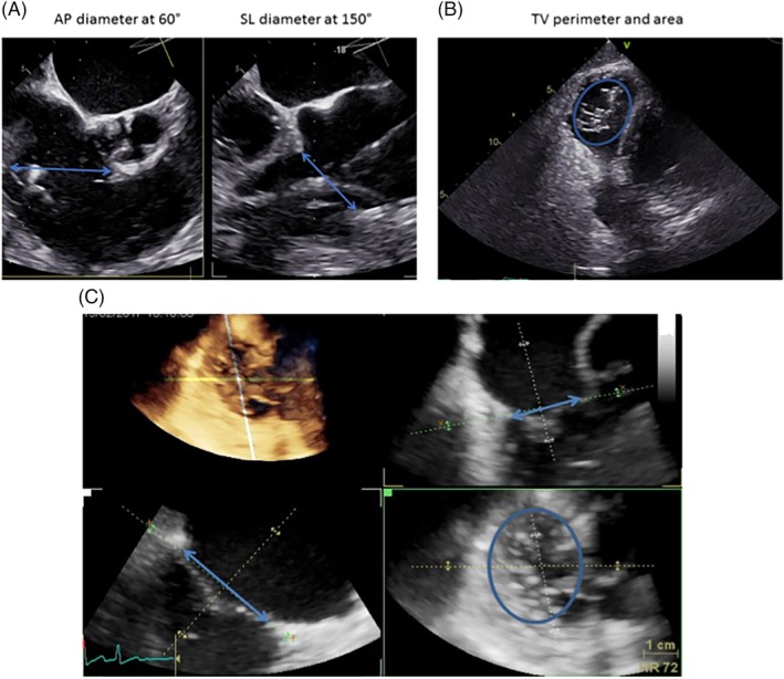
Figure 1.

Assessment of tricuspid valve (TV) geometry in 2D‐TEE and 3D‐TEE. A, Biplane grasping view from mid‐esophageal transesophageal echocardiography (TEE) to visualize anteroposterior (AP)‐ and septolateral (SL)‐diameter. B, Transgastric view at 40° to visualize TV annulus. C) Multiplane reconstruction of complete volume dataset from transgastric “en‐face” view at 20‐40° (TV view from the right atrium side): Dimensional assessment of TV
