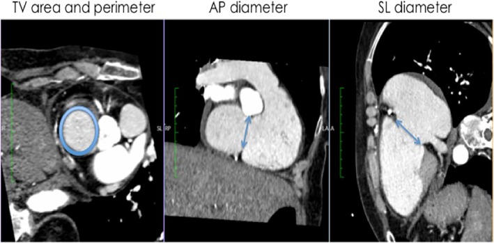
Figure 2.

Assessment of TV geometry in multislice computed tomography (MSCT). In short axis view (image on the left): tricuspid valve (TV) perimeter und cross sectional area; in two‐chamber long‐axis view (image in the middle): anteroseptal diameter; in right ventricle (RV) inflow‐outflow view (image on the right): TV septolateral diameter
