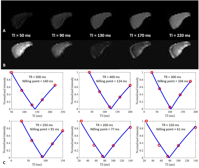
Figure 3.
A cortical bone sample from a 90-year-old donor imaged with a dual-echo 3D IR-UTE Cones sequence with a TR of 500 ms, and a TE of 32 μs (A) and 2.5 ms (B). Signal intensity for the first echo increases with TI from 50 to 220 ms (A), while signal intensity for the second echo decreases with TI until a minimum signal intensity is reached at a TI of 130 ms; it then increases with TI (B). Excellent curve fitting was achieved for IR-UTE signal intensities of the second echo at five TIs, allowing accurate estimation of pore water nulling times which were 140, 124, 104, 91, 77, and 61 ms for TRs of 500, 400, 300, 250, 200 and 150 ms, respectively (C).

