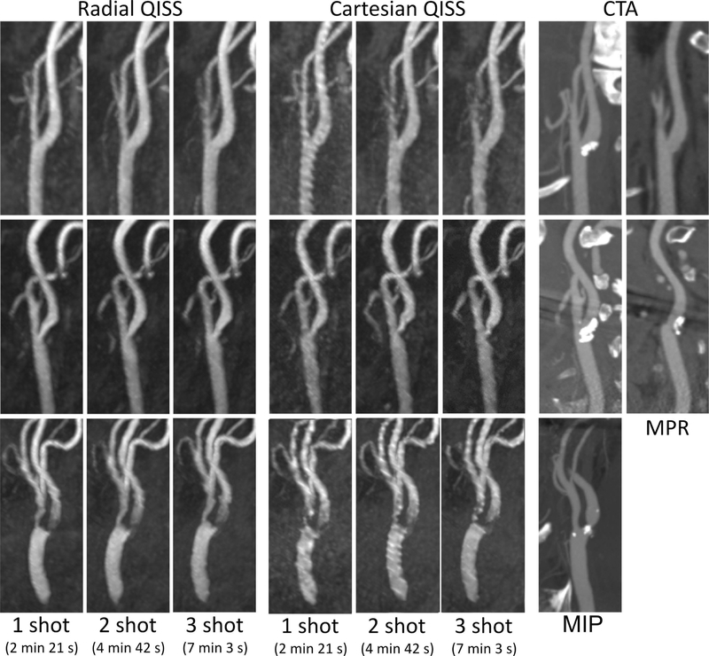
Figure 6.
Maximum intensity projection (MIP) images comparing ungated radial and Cartesian QISS obtained in three patients (top, middle, and bottom panels) with disease at the carotid bifurcation. Note the consistently improved arterial portrayal obtained with radial as opposed to Cartesian QISS, the feasibility of rapid ungated 1-shot radial QISS, and the correlation of ungated radial QISS with contrast-enhanced computed tomography angiography (CTA) obtained between 3 to 9 weeks prior. Multiplanar reformations (MPR) shown for bifurcations obscured by substantial calcification.

