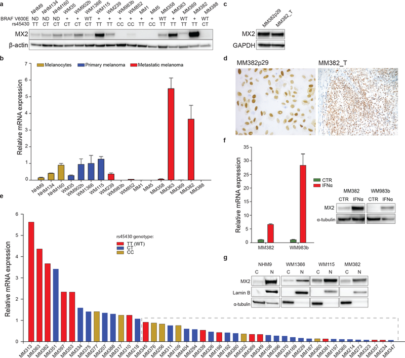
Figure 1. Characterization of MX2 expression.
a) Analysis of MX2 protein expression by immunoblotting (β actin used as a loading control). BRAF V600E and rs45430 status specified under the cell names: (ND) – not determined, (WT) – wild type, (+) – mutation is present. b) MX2 mRNA expression in normal human melanocytes (NHM), primary and metastatic melanoma lines (mRNA expression is presented as a mean value ±SE of three independent experiments). MX2 mRNA expression is normalized to primary melanoma WM1366 cell line. c) Comparison of MX2 protein expression in established melanoma WM382 line and original tumor sample by immunoblotting and d) immunohistochemistry. e) MX2 mRNA expression in metastatic melanoma tumor samples. Tumors expressing lower MX2 mRNA levels compared to primary WM1366 are inside the dashed rectangle. Columns are colored according to rs45430 genotype. f) Increase of MX2 mRNA and protein expression after treatment with IFNα 1000 IU/ml for 24 h (mRNA expression is presented as a mean value ±SE of three independent experiments). g) Cytoplasmic and nuclear expression of MX2 in normal human melanocytes, primary and metastatic melanoma cell lines examined by immunoblotting. Each MX2 blot was visualized separately.

