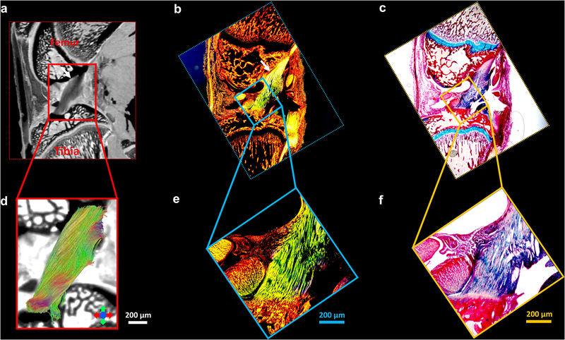
Figure 8.
The Alcian blue/Picrosirius red stain (c, f) and PLM images (b, e) of ligament, the b0 image (a), and the corresponding tractography results (d). The ligaments are indicted by the white arrows (a-c) and enlarged in Figure 8d-8f. The collagen fiber orientation in ligament is relatively uniform, which is consistent with tractography findings.

