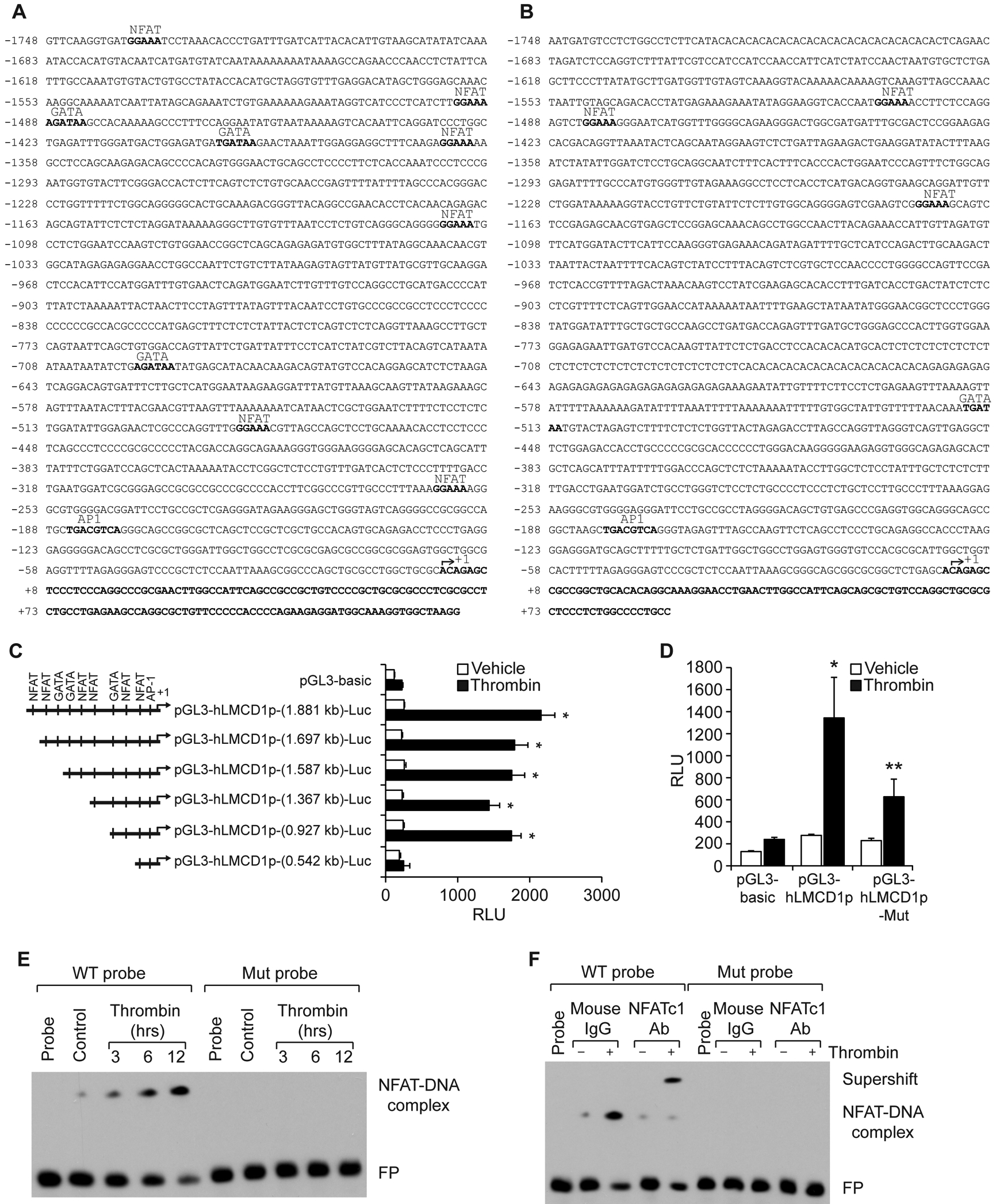
Figure 2. NFATc1 mediates LMCD1 promoter activity in HASMCs.

A & B. TRANSFAC analysis of human (A) and mouse (B) LMCD1 promoter sequence for the identification of potential transcription factor binding sites. C. HASMCs that were transfected with pGL3-basic vector or pGL3-hLMCD1 promoter-reporter gene constructs with serial 5′-deletions were quiesced and treated with and without thrombin (0.5 U/ml) for 8 hrs and analyzed for luciferase activity. D. HASMCs were transfected with pGL3-basic vector or pGL3-hLMCD1 promoter (0.927 kb) with and without the mutated NFAT-binding site (at −485 nt), quiesced, and treated with and without thrombin (0.5 U/ml) for 8 hrs, and luciferase activity was measured. E. Nuclear extracts of control and various time periods of thrombin (0.5 U/ml)-treated HASMCs were analyzed by EMSA using the NFAT-binding element at −485 nt as a biotin-labeled DNA probe. F. Nuclear extracts of control and 8 hrs of thrombin (0.5 U/ml)-treated HASMCs were analyzed for the presence of NFTAc1 in the protein-DNA complexes by supershift EMSA. The bar graphs represent Mean ± S.D. values of three independent experiments. *, p < 0.05 versus vehicle control; **, p < 0.05 versus Thrombin. RLU, relative luciferase units; FP, free probe.
