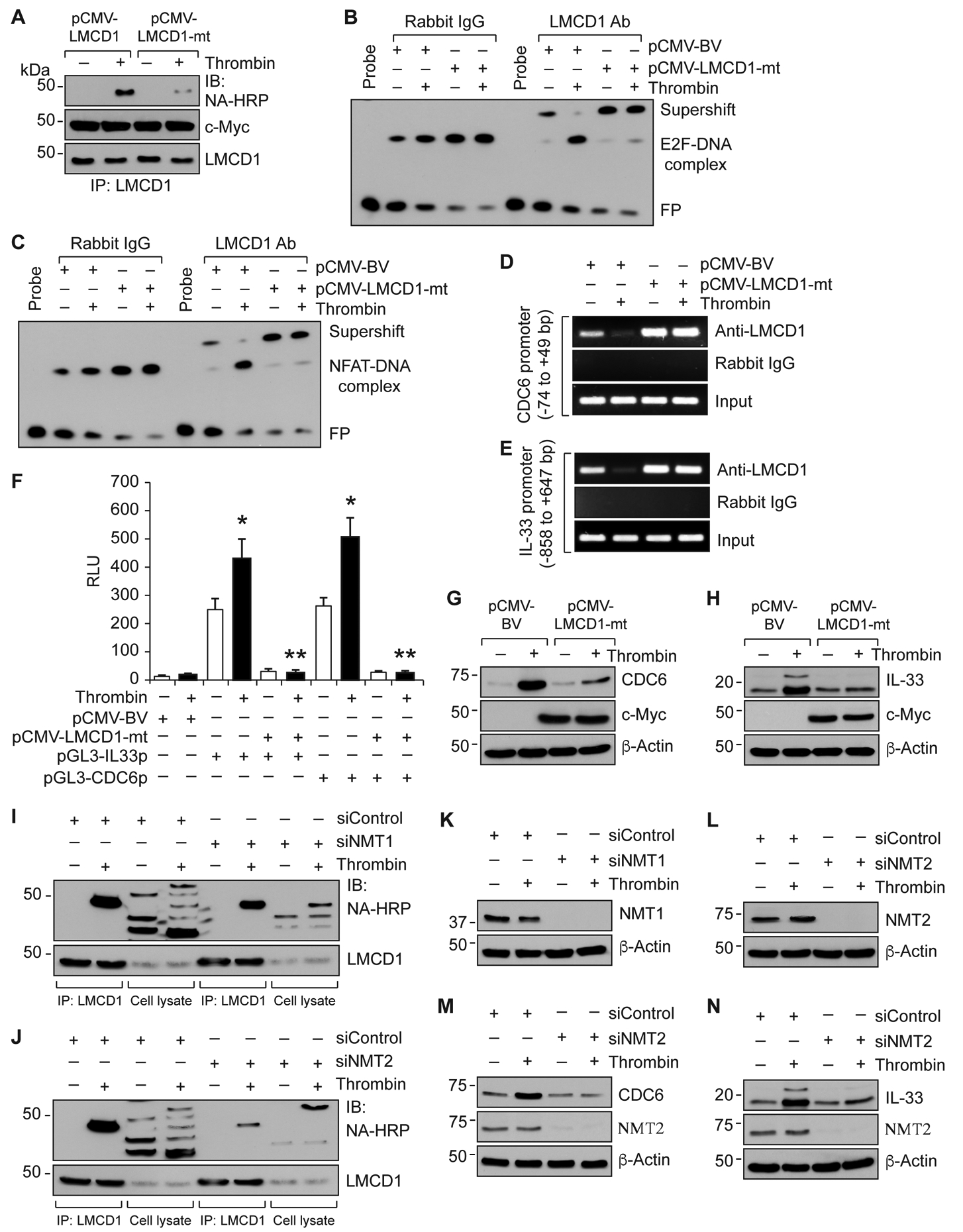
Figure 7. N-Myristoyl transferase 2 mediates thrombin-induced myristoylation and derepression of LMCD1 in the regulation of E2F1-mediated CDC6 and NFATc1-mediated IL-33 expression in MASMCs.

A. MASMCs were transfected with pCMV-LMCD1 or pCMV-LMCD1-mt vectors and 36 hrs later cells were quiesced, metabolically labeled with azidomyristate (40 μM) for 3 hrs and treated with and without thrombin (0.5 U/ml) for 8 hrs. Cell extracts were prepared, immunoprecipitated with anti-LMCD1 antibody, immunocomplexes were eluted with phosphine reaction buffer, conjugated to phosphine-biotine (250 μM) and analyzed by Western blotting using neutravidin-HRP and normalized to LMCD1 levels. B & C. MASMCs that were transfected with the indicated vectors and treated with and without thrombin (0.5 U/ml) for 8 hrs were analyzed by supershift EMSA for the presence of LMCD1 in the protein-DNA complexes of CDC6 (B) or IL-33 (C) promoters using their specific biotin-labeled probes. D & E. All the conditions were the same as in panels B & C except that cells were subjected to ChIP assay for CDC6 promoter region encompassing E2F site (D) or IL-33 promoter region encompassing NFAT site (E) using anti-LMCD1 antibodies. F. MASMCs were transfected with pCMV-basic or pCMV-LMCD1-mt vector in combination with pGL3-hIL33p or pGL3-hCDC6p and 36 hrs later cells were quiesced, treated with and without thrombin (0.5 U/ml) for 8 hrs, cell extracts were prepared and analyzed for luciferase activity. G & H. MASMCs that were transfected with pCMV-basic or pCMV-LMCD1-mt vectors were quiesced, treated with or without thrombin (0.5 U/ml) for 8 hrs, cell extracts were prepared and analyzed by Western blotting for CDC6 and IL-33 levels using their specific antibodies. The same membranes were reprobed for β-Actin for normalization. I-L. MASMCs were transfected with the indicated siRNA (100 nM) and 36 hrs later cells were quiesced, metabolically labeled with azidomyristate (40 μM) for 3 hrs and treated with and without thrombin (0.5 U/ml) for 8 hrs. Cell extracts were prepared, immunoprecipitated with anti-LMCD1 antibody, immunocomplexes were eluted with phosphine reaction buffer, conjugated to phosphine-biotine (250 μM) and analyzed by Western blotting using neutravidin-HRP and normalized to LMCD1 levels (I & J). Cell extracts were also analyzed for siRNA efficacy (K & L). M & N. MASMCs that were transfected with siNMT2 (100 nM) were quiesced, treated with or without thrombin (0.5 U/ml) for 8 hrs, cell extracts were prepared and analyzed by Western blotting for CDC6 and IL-33 levels. The membranes were reprobed for NMT2 and β-Actin. The bar graphs represent Mean ± S.D. values of three independent experiments. *, p < 0.05 versus pGL3-basic vector; **, p < 0.05 versus pGL3-hCDC6p or pGL3-hIL33p. NA-HRP, neutravidin-HRP; NMT1, N-Myristoyltransferase 1; NMT2, N-Myristoyltransferase 2.
