
Figure 8. Differential expression of LMCD1 in human and mouse neointimal hyperplasia.
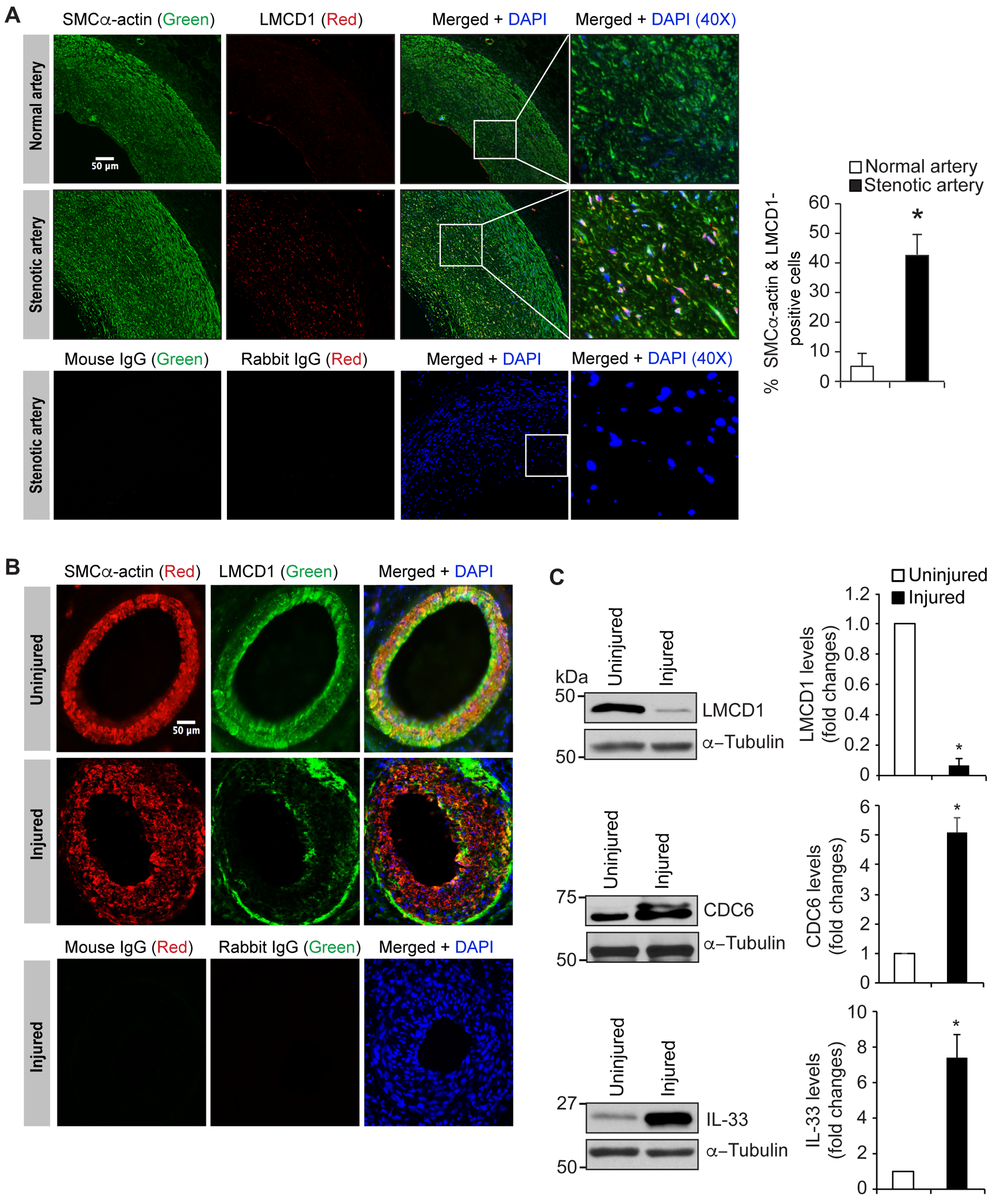
A. Human stenotic and non-stenotic artery sections were stained for SMCα-actin (green) and LMCD1 (red). The sections were also stained with normal mouse or rabbit IgG as negative controls. B. Uninjured and 3 weeks of post guidewire-injured mouse femoral artery cross-sections were stained for SMCα-actin (red) and LMCD1 (green). C. Tissue extracts prepared from uninjured and guide wire-injured mouse femoral arteries were analyzed by Western blotting for LMCD1, CDC6 and IL-33 levels using their specific antibodies and normalized to α-tubulin levels. The bar graphs represent Mean ± S.D. values of six animals (two pooled arteries for each Western blotting analysis). *, p < 0.05 versus uninjured. Scale bar is 50 μm.