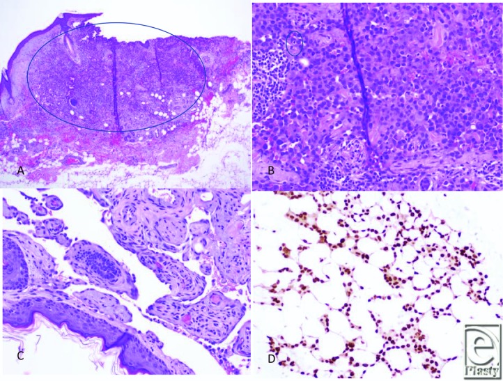
Figure 2.

Histologic images of cutaneous angiosarcoma. (a) Left scalp excision revealed multiple foci of malignant cells involving the dermis and superficially involving subcutaneous soft tissue. Note this area demonstrates a solid area of growth of epithelioid cells. (H&E, ×4). (b) Areas of the tumor reveal malignant epithelioid cells with vague rudimentary vasoformation. The epithelioid cells exhibit round/ovoid irregular nuclei with abundant cytoplasm. Mitotic figure is noted (H&E, ×20). (c) Other areas of the tumor demonstrate anastomosing vessels lined by plump, hyperchromatic endothelial cells with focal papillations (H&E, ×20). (d) The tumor shows vasoformative growth within subcutaneous tissue. Tumor cells are positive for ERG expression (Immunostain, ×20). HE indicates hematoxylin and eosin.
