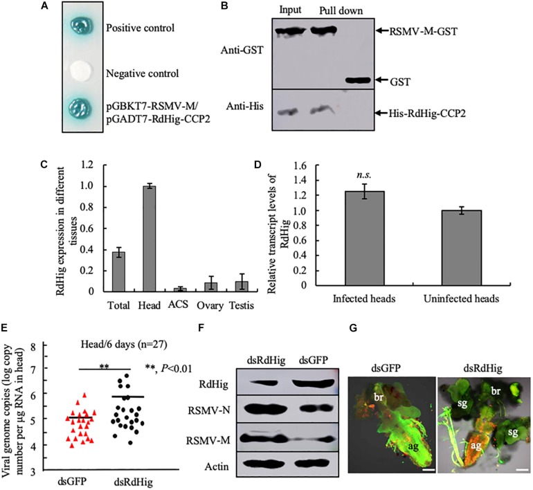
FIGURE 5.
RdHig has antiviral activity during RSMV infection of the CNS of R. dorsalis. (A) Yeast two-hybrid assay to detect the interaction of RSMV M protein with the CCP2 domains of RdHig. (B) GST pull-down assay to verify the interaction of RdHig CCP2 domains with RSMV M protein. GST protein was used as the control. (C) RT-qPCR assay to detect RdHig transcript levels in different tissues of R. dorsalis. ACS, alimentary canal. Values are the means (±SE) of three biological replicates. (D) The transcript levels of RdHig expression in RSMV-infected or uninfected heads of R. dorsalis, as detected by an RT-qPCR assay. Values are the means (±SE) of three biological replicates. (E) RT-qPCR assay of viral genome copies of RSMV in the heads of R. dorsalis at 6 days post-microinjection of dsRNAs. RSMV genome copies in the individual heads (n = 27) of viruliferous R. dorsalis were calculated as the log of the copies number/μg RNA in head based on the standard curve for the RSMV N gene. P-values were estimated using a Student’s t-test. (F) The accumulation of RdHig, RSMV N, and RSMV M in the heads of viruliferous R. dorsalis at 6 days post-microinjection of dsRdHig and dsGFP, as detected by an immunoblot assay. Insect β-actin was used as the internal control. (G) RSMV infection of the CNS of viruliferous R. dorsalis at 6 days post dsRNA microinjection, as analyzed by an immunofluorescence assay. Virus-infected heads were immunolabeled with RSMV-M-rhodamine (red) and α-tubulin-FITC (green). ag, abdominal ganglion; br, brain; sg, salivary gland. Bars, 50 μm.

