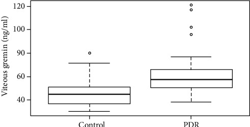
Figure 1.

The vitreous concentration of gremlin in patients with PDR and idiopathic epimacular membrane. Box plots showed that the vitreous concentration of gremlin in patients with PDR was significantly higher than in patients with idiopathic epimacular membrane (p < 0.001). PDR represents proliferative diabetic retinopathy; control represents idiopathic epimacular membrane.
