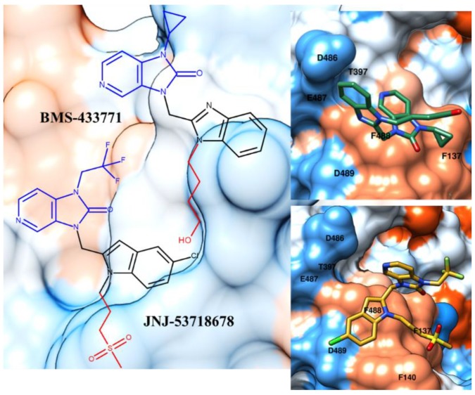
Figure 4.
Chemical structure and X-ray positioning of BMS-433771 (pdb code: 5EA7) [31] and JNJ-53718678 (pdb code: 5KWW) [32] in complex with the RSV F protein. The chemical motifs of the two inhibitors featuring quite comparable contacts with the biological target are highlighted in blue and red. Hydrophobic and polar areas of the protein are represented as blue and orange regions on the RSV F protein’s Connolly surface.

