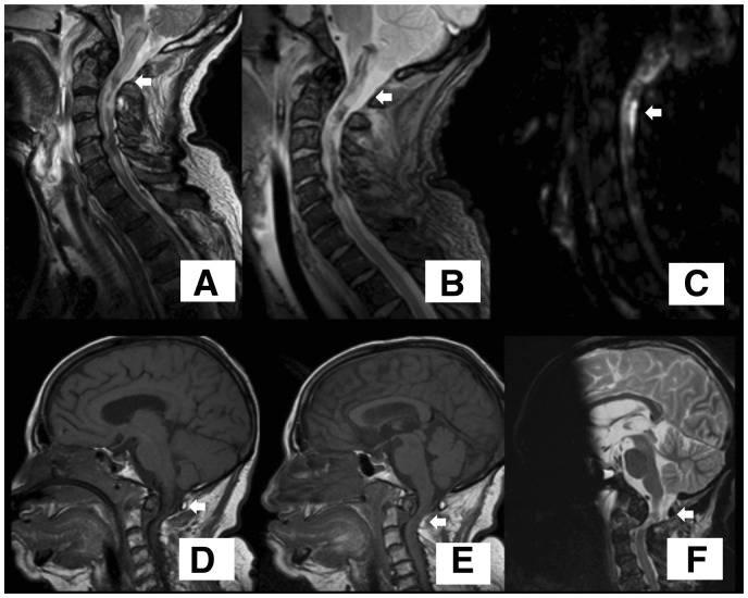
Figure 3.
Cervical cord magnetic resonance imaging. Odontoid fracture with luxation D’Alonso type II with secondary spinal cord injury due to compression (arrows). (A–C) Cervical magnetic resonance imaging of Patient 1. (A) FLAIR sequence. (B) T2 sequence. (C) DWI sequence. (D–F) Craniocervical magnetic resonance imaging of Patient 2. (D and E) T1 sequence of Patient 2. (F) T2 sequence.

