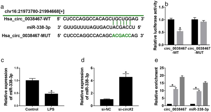
Figure 4.

Circ_0038467 acted as a sponge of miR‐338‐3p in 16HBE cells. (a) Schematic model of the miR‐338‐3p‐binding sites within circ_0038467 identified by the online database Circinteractome and the mutant in the target sequence. (b) Luciferase activity was monitored in 16HBE cells cotransfected with circ_0038467 wild‐type reporter construct (circ_0038467‐WT) or circ_0038467 mutant‐type construct (circ_0038467‐MUT) and miR‐338‐3p mimic or miR‐NC mimic.  miR‐NC,
  miR‐338‐3p. (c) MiR‐338‐3p expression was detected by qRT‐PCR in 16HBE cells after LPS treatment. (d) MiR‐338‐3p expression was determined in 16HBE cells transfected with si‐NC or si‐circ#2. si‐circ#2: si‐circ_0038467#2. (e) Cell lysates were incubated with anti‐Ago2 or anti‐IgG antibody, and enrichment of circ_0038467 and miR‐338‐3p were evaluated by qRT‐PCR. *P < 0.05.
