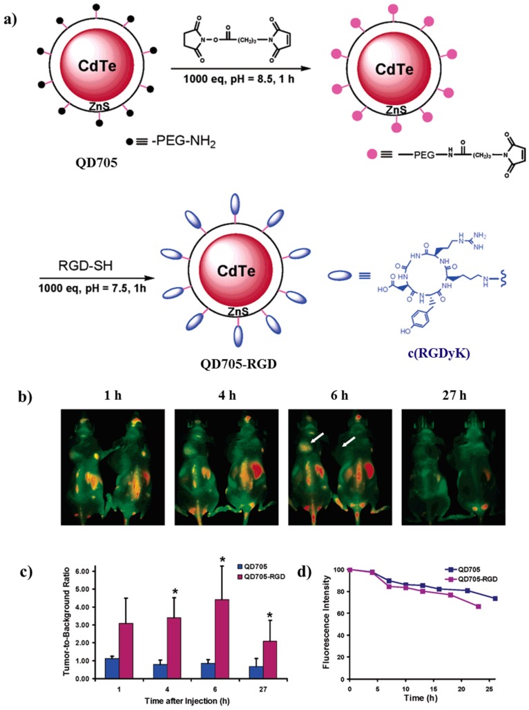
Figure 14.
(a) Synthetic pathway for the preparation of QD705-RGD. (b) In vivo NIR fluorescence imaging of U87MG tumor-bearing mice (left shoulder, pointed by white arrows) injected with 200 pmol of QD705-RGD (left) and QD705 (right), respectively. All images were acquired under the same instrumental conditions. The mice autofluorescence is color coded green while the unmixed QD signal is color coded red. Prominent uptake in the liver, bone marrow, and lymph nodes was also visible. (c) Tumor-to-background ratios of mice injected with QD705 or QD705-RGD. The data were represented as mean (standard deviation (SD)). Using one-tailed paired Student’s t-test (n) 3),”*” denotes where p < 0.05 as compared to the mice injected with QD705. (d) Serum stability of QD705 and QD705-RGD in complete mouse serum over the course of 24 h. Reprinted with permission from [95]; copyright (2006) American Chemical Society.

