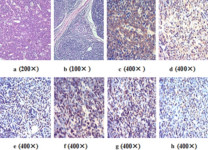
Figure 3.

(a) On microscopic visualization, the tumour had a highly vascular appearance, magnification 200x. (b) Some areas were type B1 thymoma (lymphocyte‐rich thymoma), and a few neoplastic epithelial cells could be seen against a background of lymphocytes, magnification 100x. (c) CK19 was positive for epithelial cells, magnification 400x. (d) CD99 was positive for lymphocytes, magnification 400x. (e) TdT was positive for lymphocytes, magnification 400x. (f) CD5 was positive for lymphocytes, magnification 400x. (g) CD3 was positive for lymphocytes, magnification 400x. (h) Ki67 was positive for lymphocytes, magnification 400x.
