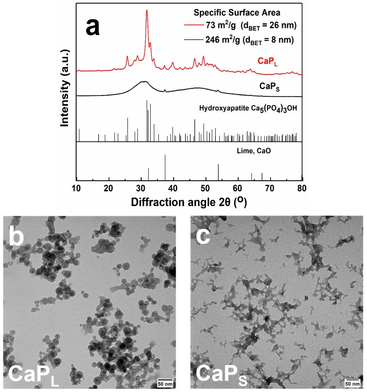
Figure 1.
(a) X-ray diffraction (XRD) patterns of the CaP nanoparticles. The specific surface area (SSA), as determined by the nitrogen adsorption–desorption isotherms, along with the corresponding primary particle size, dBET, are also shown. By varying FSP synthesis conditions either crystalline or amorphous particles are obtained. Main peaks are assigned to hydroxyapatite, Ca5(PO4)3OH, whereas CaO is also observed. Transmission electron microscopy (TEM) images of as-prepared CaPL (b) and CaPS (c) samples. CaPL particles are spherical with loosely agglomerated structure, while fused particles with sintered necks are clearly illustrated for CaPS (Scale bar 50 nm).

