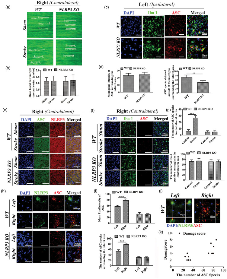
Figure 5.
Expression of NLRP3/ASC in the ipsilateral and contralateral cortices of WT and NLRP3 KO mice following single left photothrombotic stroke or bilateral stroke. (a) XY scanning of target vessels in the right cortex following single left photothrombotic stroke. (b) Comparisons of the blood flows in the right cortex. (c) Immunofluorescence staining of ASC and Iba-1-positive microglia. (d) Comparison of the ASC pixel intensity and the number of ASC specks surrounding the stroke area between WT and NLRP3 KO mice following single left photothrombotic stroke. (e–f) Immunofluorescence staining of ASC and NLRP3(E), ASC and Iba-1-positive microglia (f) in the right (contralateral) cortex following single left photothrombotic stroke. (g) Comparison of ASC specks and Iba-1-positive microglia in the right parietal cortex between WT and NLRP3 KO mice with or without single left photothrombotic stroke. (h) Immunofluorescence staining of ASC and NLRP3 surrounding the first (left) and recurrent (right) parietal infarct areas in WT and NLRP3 KO mice following bilateral stroke at interval of one week. (i) Comparisons of the pixel intensity and the number of ASC specks surrounding the infarct area following bilateral stroke. (j) Co-localization of ASC and NLRP3 in WT mice following bilateral stroke. (k) Pearson correlation analysis about the number of ASC specks surrounding the second infarct area and the damage score of the second stroke *P ≤ 0.05; **P ≤ 0.01; ***P ≤ 0.001. n = 6.

