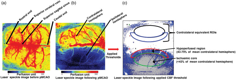
Figure 1.
Laser speckle contrast imaging set up and analysis. (a) A representative laser speckle image showing normally perfused rat brain with superior cortical vessels before pMCAO was induced. (b) A representative laser speckle image of the cortical surface following pMCAO, showing normal blood flow in the contralateral hemisphere (red–yellow) and cortical blood flow deficit in the ipsilateral hemisphere (blue–black). (c) ROIs on the LSCI defined with applied CBF thresholds: ischaemic core (cortical blood flow < 43% of mean contralateral hemisphere), hypoperfused tissue (cortical blood flow between 43 and 75% of mean contralateral hemisphere) along with contralateral equivalent ROIs.

