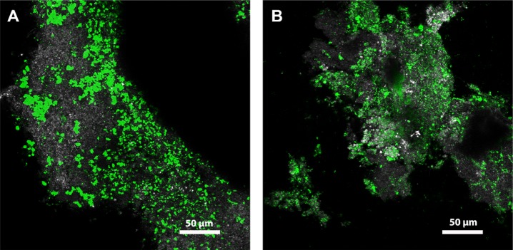
Figure 2.
Confocal laser scanning microscopy of fully hydrated, intact anammox granules stained with fluorescently labeled lectins shown as maximum intensity projections. (A) WGA (Wheat germ agglutinin, 40 optical sections) and (B) HAA (Helix aspersa agglutinin, 66 optical sections). Color allocation: grey: reflection signal, green: lectin staining.

