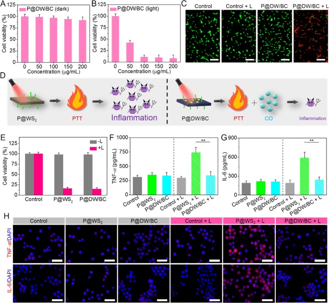
Figure 4.
Cell viability of CT26 cells treated with P@DW/BC NSs with various concentrations (A) in the dark condition and (B) under 808 nm laser irradiation (1 W cm–2, 6 min). (C) Live/dead staining images of CT26 cells after various treatments. Scale bar: 100 μm. Viable cells were stained green with calcein-AM, and dead/late apoptosis cells were stained red with PI. (D) Schematic of the different results after P@WS2- or P@DW/BC NSs-mediated PTT. P@WS2-mediated PTT was supposed to induce obvious inflammation, while the inflammatory reactions induced by P@DW/BC-mediated PTT was supposed to be largely reduced due to the generation of CO. (E) Cell viability of CT26 cells after various treatments. (F) TNF-α and (G) IL-6 levels of RAW 264.7 macrophages after treatment with media of different CT26 cell samples, n = 3, **p < 0.01. (H) Representative immunofluorescence images of TNF-α and IL-6 expression in RAW 264.7 macrophages after treatment with media of different CT26 cell samples. Scale bar: 100 μm.

