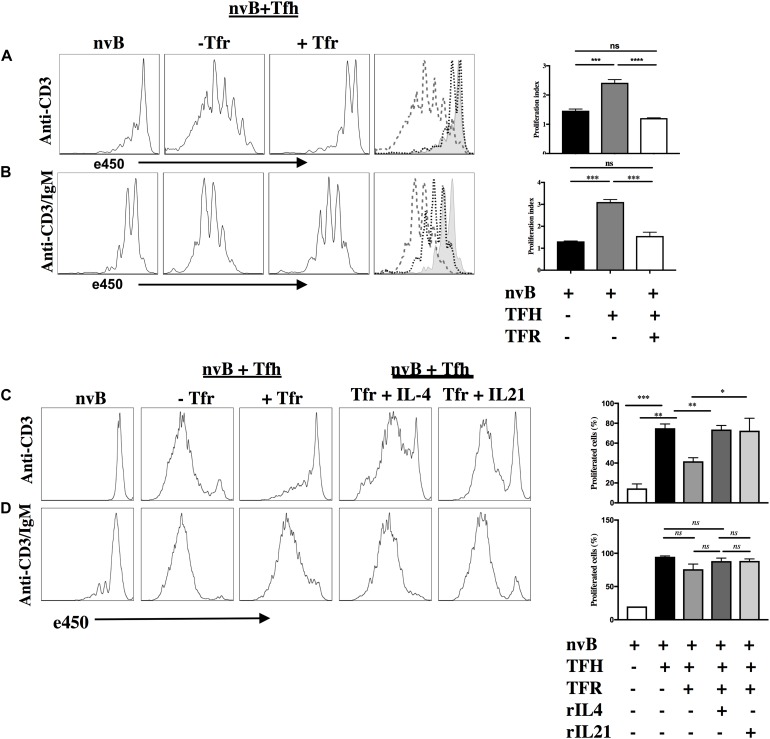
FIGURE 4.
Tfr cells suppress B cell proliferation in a T-cell dependent manner. (A) nvB were labeled with eF450 and cultured with primed Tfh cells (dashed), anti-CD3, in the presence or absence of primed Tfrs (dotted). (B) e450-labeled nvB cells were cultured with primed Tfh cells, stimulated with anti-CD3/anti-IgM in the presence of absence of Tfr cells. (C) As in panel (A), except IL-4 or IL-21 were added to cultures with Tfr cells. (D) As in panel (B) except cultures with Tfr cells were supplemented with IL-4 or IL-21. Control B cells were stimulated with anti-IgM (5 μg/ml) alone. Statistical results are from 10 independent experiments showing SEM (n = 10). *p < 0.05, **p < 0.005, ***p < 0.0001, ****p < 0.00001, ns: not significant; unpaired Student t-test.

