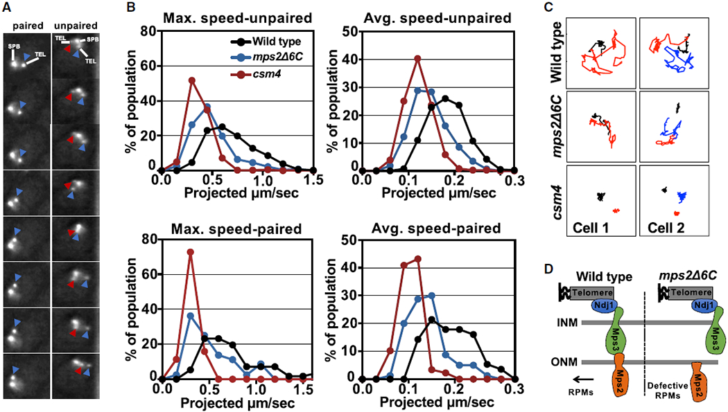
Figure 2. Mps2 Promotes Rapid-Prophase Movements in Budding Yeast.

(A) The image frames in the pictures provide an illustration of how speed was measured in cells with paired (left) and unpaired (right) chromosomes 4 right telomeres. These images were acquired with through-focus microscopy, every 1 s, for a total of 61 frames (detailed description for through-focus microscopy and its quantification and semi-auto analyses were published in [16]; see Video S1). In different frames, the same color arrows mark the same lacO256/lacI-GFP spots.
(B) Histograms display meiotic rapid-prophase movements activity in zygotene by measures of maximum and average speed for unpaired and paired chromosomes (see Figure S2) in mps2Δ6C mutant, wild-type, and csm4Δ strains. In our strain background, for strains that are not delayed in meiotic progression, 4 h post-shift into sporulation medium roughly correlates with zygotene and 5 h post-shift correlates with pachytene (see Figure S3). For csm4Δ, a mutant known for prolonged delay, 4.5 h post-shift roughly correlates with zygotene [16], is used as a control for near absence of rapid-prophase movements. All measurements are for lacO256/lacI-GFP spots adjacent to the 4R telomere, and each profile is the result of a total of three independent experiments (250–300 cells scored per strain), where there are 2 spots (unpaired, top) or 1 spot (paired, lower panel).
(C) Example of traces (spots in 61-frame time-lapse series) of paired and unpaired telomere movement in wild-type, mps2Δ6C, and csm4Δ in zygotene cells. (Black line marks the spindle pole body movement. Figures with red line only mark paired telomere and with both red and blue lines mark unpaired telomeres.) Represented are projection images of telomere movement in 61 s. Cell 1 and cell 2 are examples of cells with paired and unpaired telomeres, respectively.
(D) Schematic of wild-type Mps2 and Mps2Δ6C association to other components of the LINC complex.
