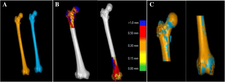
Fig. 2.
Stages of volume fusion of the proximal and distal femur from (a) the original right femur and the mirrored left femur. b The color scale indicates how closely the software could place the object of interest in the two CT stacks for the proximal part and the distal part, respectively. c The proximal femur is assumed as the static portion (reference) to report rotation and translation of the distal femur

