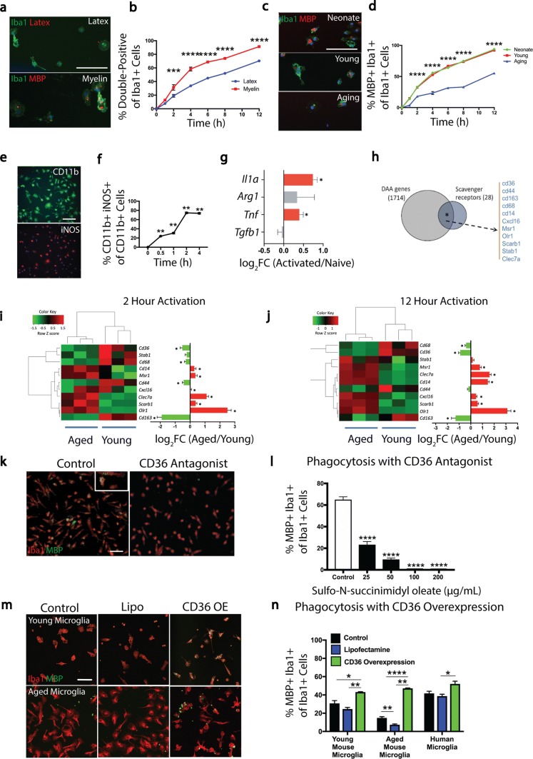
Fig. 1.
Myelin debris activates microglia and myelin phagocytosis requires the scavenger receptor CD36. a Representative images of mouse microglia exposed to myelin debris and fluorescently conjugated latex beads (red) and stained for Iba1 (green) and MBP (red). b A greater percentage of microglia phagocytose myelin debris compared to latex beads over time. c Representative images of neonate, young adult, and aged microglia exposed to myelin debris and stained for Iba1 (green) and MBP (red). d Neonate and young microglia phagocytose myelin debris with a significantly greater efficiency than aged microglia. e Representative images of mouse microglia exposed to myelin debris and stained for CD11b (green) and iNOS (red). f Myelin debris stimulates the expression of iNOS in CD11b+ microglia. g Log2 fold change of RNA expression of pro- and anti-inflammatory genes from microglia activated with myelin debris for 2 h compared to naïve controls. h Venn diagram representation of the association of scavenger receptors in the set of differentially expressed genes (DAA) whose expression in microglia was regulated upon activation and that is affected due to aging. i, j Heat maps of expression levels of scavenger receptors (in DAA) in microglia isolated from young and aging mice and activated for 2 (i) and 12 h (j). Euclidean distance matrix was used for hierarchical clustering. Difference in expression of scavenger receptors in microglia from aged mice was compared to microglia from young mice and the estimated log2 fold change was plotted. k Representative images of mouse microglia exposed to myelin debris and treated with a CD36 receptor antagonist (sulfo-N-succinimidyl oleate, SSO) and stained for Iba1 (red) and MBP (green). l SSO reduces microglia phagocytosis of myelin debris. m Representative images of young and aged microglia exposed to myelin debris and treated with control, lipofectamine (Lipo), and overexpression of CD36 (CD36 OE) and stained for Iba1 (red) and MBP (green). n Overexpression of CD36 significantly enhances myelin phagocytosis by young and aged mouse microglia as well as human microglia. Values are represented as mean with the standard error of the mean. Results were analyzed with a two-way repeated-measures ANOVA with a Bonferroni’s post hoc test (b, d), a one-way repeated-measures ANOVA with a Dunnett’s post hoc test (f, l), a Wald test and Benjamin–Hochberg p value adjustment (g, i, j), a two-sided Chi-square test with Yates’ correction (h), and a one-way ANOVA with a Bonferroni’s post hoc test (n each cell type analyzed individually). Values are indicative of triplicate cultures. For panels a, c, e, k, m, scale bars equal 50 μm. For panels b, d, f, l, n, *p < 0.05; **p < 0.01; ***p < 0.001; ****p < 0.0001. For panels g, i, j, *p < 0.05

