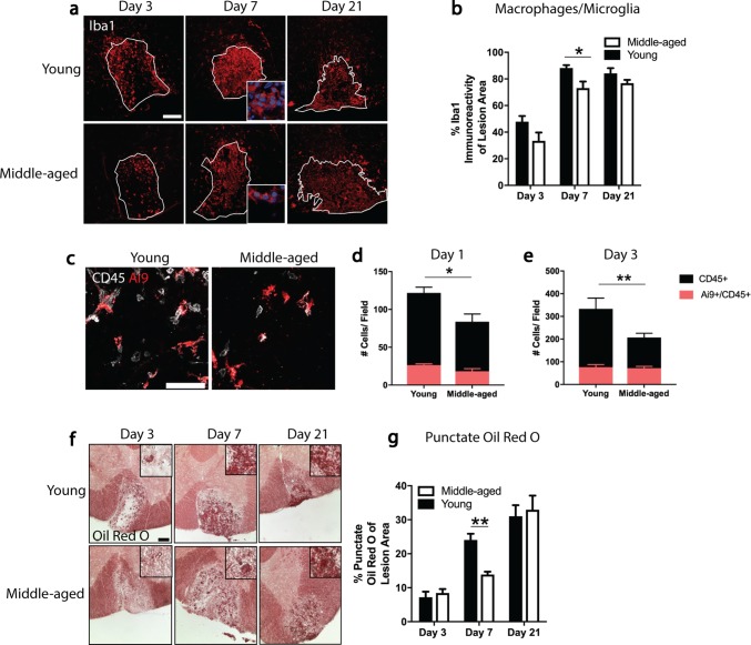
Fig. 2.
Middle-aged mice have decreased monocyte-derived macrophage recruitment and impaired phagocytosis. a Representative images of lesions from young (2–3 month) and middle-aged (9–12 month) mice stained with the macrophage/microglia marker-ionized calcium-binding adaptor molecule-1 (Iba1) at 3, 7, and 21 days post-demyelination. b Lesions in middle-aged mice have significantly less Iba1 immunoreactivity than lesions in young mice at 7 days. c Representative images of lysolecithin lesions from young and middle-aged CX3CR1CreER:Rosa26TdT (Ai9) mice stained with an antibody against CD45. d, e The number of microglia (Ai9+/CD45+) within the lesion does not differ between young and middle-aged mice. The number of peripheral leukocytes (CD45+), however, is significantly less in middle-aged mice at both time points. f Representative bright field images of lesion epicenters from young and middle-aged mice stained with Oil Red O (ORO) at 3, 7, and 21 days post-demyelination. g Lesions in young mice have significantly more punctate ORO staining than lesions in middle-aged mice at 7 days post-demyelination. Values are represented as mean with the standard error of the mean. Results were analyzed with a two-way ANOVA with a Bonferroni’s post hoc test (b, d, e, g). For panels b, d, e, and g, between five and nine mice were analyzed per group. For panels a and f, scale bars equal 100 μm. For panel c, scale bars equal 50 μm. *p < 0.05; **p < 0.01

