Fig. 5.
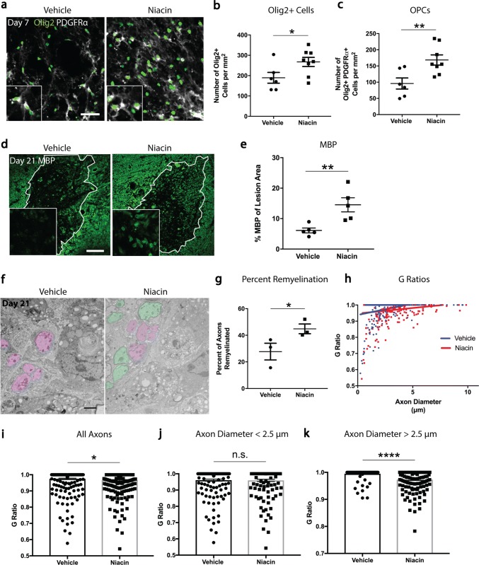
Niacin treatment of middle-aged mice enhances recruitment of OPCs and rejuvenates remyelination. a Representative images of lesion epicenters double-stained with Olig2 (green) and PDGFRα (white) from vehicle- and niacin-treated mice at 7 days post-demyelination. b, c Seven day lesions in niacin-treated mice have a significant increase in the densities of Olig2+ oligodendrocyte lineage cells and Olig2+PDGFRα+OPCs. d Representative images of lesion epicenters stained for MBP (green) from vehicle- and niacin-treated mice at 21 days post-demyelination. MBP-positive myelin rings (with zoomed images as inserts) are low in intensity whereas MBP-positive debris is high in intensity. e There is a significant increase in the percentage of MBP-positive area within the lesions of niacin-treated mice. f Representative electron micrographs of day 21 lesions from middle-aged mice treated with either vehicle or niacin. Demyelinated axons are depicted as magenta whereas remyelinated axons are shown in green.g Lesion cores from middle-aged mice treated with niacin have a significant increase in the percentage of axons that are remyelinated compared to lesions from vehicle-treated mice. h Graph displaying g ratios as a function of axon diameter from mice treated with either vehicle or niacin. i Lesions from middle-aged mice treated with niacin have a significant decrease in the mean g ratio (i.e. more remyelinated axons) across all axons analyzed. j Small caliber axons (diameter < 2.5 μm) do not differ in g ratio between vehicle and niacin treatment. k Lesions from middle-aged mice treated with niacin have significantly decreased mean g ratio of large caliber axons (diameter > 2.5 μm). Values are represented as mean with the standard error of the mean. For panels b, c, e, g, i, j, and k, results were analyzed with a one-tail student’s t test. For panels b, c, e, g, each data point represents 1 mouse. *p < 0.05; **p < 0.01; ****p < 0.0001; n.s. not significant. Scale bar equals 50 μm (a), 100 μm (d) and 2 μm (f)

