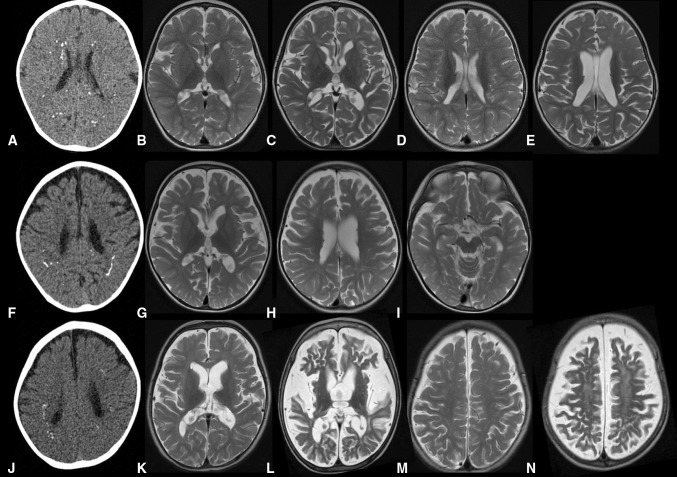
Fig. 1.
Neuroimaging of patients from F1442 and F2382. CT images of patient 1 (P1) aged 9 months (a), patient 2 (P2) aged 17 months (f), and patient 3 (P3) aged 14 months (j) demonstrate widespread spot and linear calcification in the deep and sub-cortical white matter. T2-weighted axial MR images of P1 aged 9 months (b, d) and 27 months (c, e), P2 aged 17 months (g, h, i), and P3 aged 14 months (k, m) and 27 months (l, n). Initial imaging shows mild cerebral volume loss with relatively good myelination. Follow-up shows rapidly progressive cortical and sub-cortical atrophy in P1 and P3. There is diffuse high signal in the white matter in P3 (l, n), but not P1

