Fig. 4.
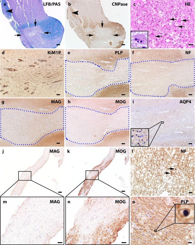
White matter pathology of MOG Ab positive inflammatory demyelinating disease. a LFB/PAS stain and myelin protein CNPase immunohistochemistry (b) on consecutive sections indicate the perivenous (arrowhead) and confluent (arrows) demyelination that coexists in the subcortical white matter of a MOGAD biopsy. c H&E stain shows marked hypertrophic reactive astrocytes present in the white matter lesion and Creutzfeldt-Peters cells are occasionally noted (inset). d–i Consecutive sections: d KiM1P immunohistochemistry indicates extensive microglia/macrophage infiltration in the white matter with no obvious border. The blue dotted lines contour the demyelinating lesion e with relative preserved axons f. The loss of minor myelin protein MAG (g) and MOG (h) are equal. AQP4 is preserved in the lesion i. j and k consecutive sections. Preferential MAG loss (j, m) with relative MOG preservation (k, n) is seen in a single MOGAD case. l Mild axonal damage characterized by axonal spheroid (indicated with arrows) is present in the demyelinating lesions. o Apoptotic oligodendrocytes with condensed nucleus (highlighted in the inset) are seen in the lesions. Scale bars in a, b, d–k = 200 μm. Scale bars in c, l and o = 20 μm

