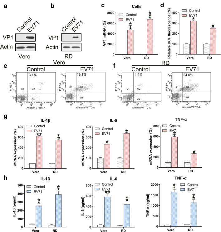
Fig. 1.
EV71 infection induces ROS generation, apoptosis, and inflammation of infected cells. a–c Reinforced mRNA and protein of VP1 in EV71-infected Vero and RD cells was identified in comparison to non-infected cells (Control) using western blotting and qPCR analyses. d ROS generation was examined in infected Vero and RD cells as evidenced by DCF fluorescence intensity assay compared to non-infected normal mice (Control). e, f Annexin V-FITC and PI flow cytometry was performed to assess the number of apoptotic Vero and RD cells. The upper right quadrant of every plot represents early dead cells. g qPCR analyses of the inflammation-promoting cytokines IL-1β, IL-6, and TNFα produced by infected cells. h The protein expression levels of IL-1β, IL-6, and TNFα of the infected cell were quantified using ELISA. Data are presented as mean ± SD. *P < 0.05, **P < 0.01, ***P < 0.001 vs. Control group

