Figure 2.
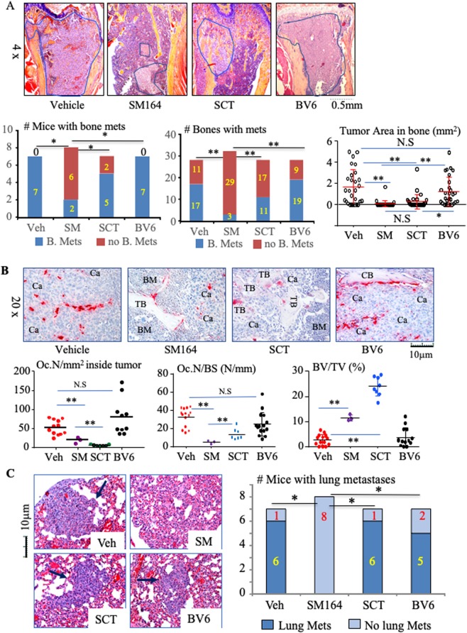
SM-164 prevents the establishment and progression of early-stage metastasis of breast cancer cells in bone and lungs. Mice, as in Fig. 1, were euthanized on day 29. (A) Representative H&E-stained sections of upper tibiae (upper panel) with tumor metastases outlined in blue. The frequency of bone metastasis, based on the number of mice (left of lower panel) and number of tibiae and femora (middle of lower panel) with bone metastases (mets), *p < 0.05 and **p < 0.01, non-parameter analysis. Tumor burden in bone, evaluated as tumor area in each long bone of the legs. *p < 0.05 and **p < 0.01, one-way ANOVA +/Dunnett test. (B) Representative TRAP-stained sections of tibiae (upper panel) to illustrate OCs (red staining) in metastatic tumor deposits. OC numbers inside metastatic cancer (left lower panel) and on trabecular bone surfaces (middle lower panel) as well as volume of non-resorbed trabecular bone (right lower panel). Ca = cancer, BM = normal bone marrow, CB = cortical bone, TB = trabecular bone. *p < 0.05 and **p < 0.01, one-way ANOVA +/Dunnett test. (C) Representative H&E-stained sections of lungs, with lung metastases arrowed, and analysis of the numbers of mice with lung metastases. *p < 0.05, non-parametric analysis.

