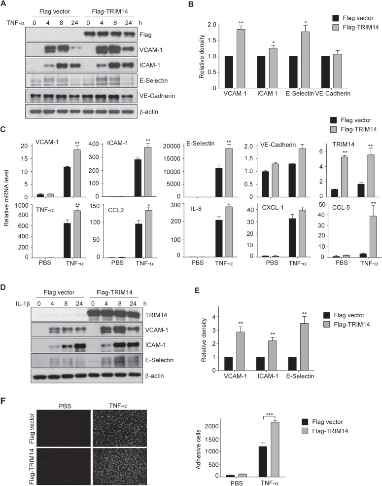
Figure 2.

Overexpression of TRIM14 increased the expression of adhesion molecules and cytokines and monocyte adherence to HUVEC. (A) HUVECs were transiently transfected with Flag-TRIM14 or empty vector for 24 h and then treated with 10 ng/ml TNF-α for 0, 4, 8, and 24 h. Cell lysates were extracted and used to detect the protein levels of VCAM-1, ICAM-1, E-selectin, and VE-cadherin. (B) Relative fold changes of proteins after 8 h treatment of TNF-α were determined by densitometry and normalized to β-actin. Data are presented as mean ± SD (n = 3); *P < 0.05, **P < 0.01 by Student’s t-test. (C) HUVECs were transfected with Flag-TRIM14 or empty vector for 24 h. Transfected cells were then stimulated with 10 ng/ml TNF-α or PBS (as controls) for 4 h. The mRNA levels of adhesive molecules and cytokines were determined by qPCR and normalized to β-actin. (D) Flag-TRIM14/Flag was transfected into HUVECs, and the transfected cells were treated with 10 ng/ml IL-1β for 0, 4, 8, and 24 h. The protein levels were detected by western blot analysis. (E) Relative fold changes of proteins after 8 h treatment of IL-1β were determined by densitometry and normalized to β-actin. Data are presented as mean ± SD (n = 3); **P < 0.01 by Student’s t-test. (F) Flag-TRIM14 or empty vector were transfected into HUVECs, and the transfected cells were incubated with TNF-α or PBS for 8 h and then co-cultured with fluorescence-labeled THP-1 cells for 1 h. After carefully washing, adhesive cells were visualized by Cytation 3 Cell Imaging Multi-Reader (Biotek Instruments). Attached cells were counted from 5 random pictures in three independent experiments. ***P < 0.001.
