Figure 5.

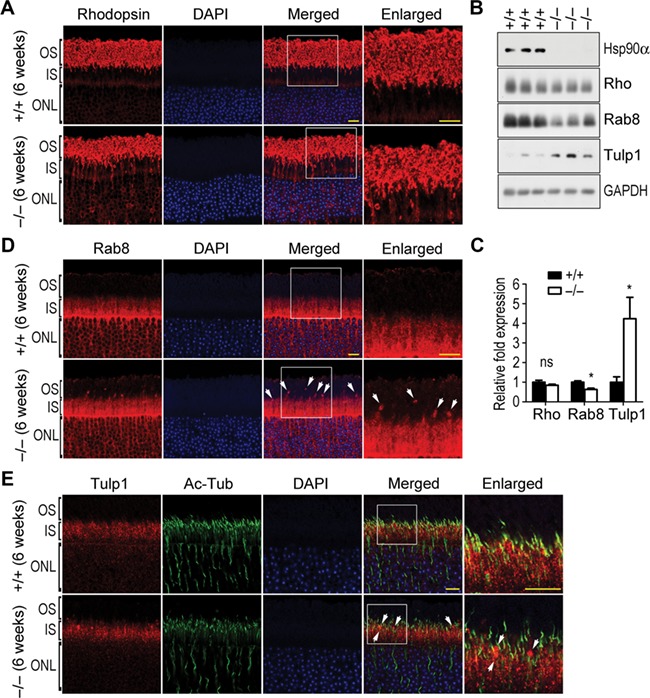
Abnormal rhodopsin transport in Hsp90α-deficient mice. Scale bar, 10 μm. The boxed areas are enlarged to show details. (A) The retention of rhodopsin in IS and ONL of photoreceptors in 6-week Hsp90α-deficient mice. (B) The expression of rhodopsin (Rho), Rab8, and Tulp1 in the retinas of Hsp90α-deficient mice or wild-type littermates. GAPDH was the protein loading control. (C) Quantification of protein expression. Rhodopsin, Rab8, and Tulp1 on western blot were subjected to densitometry analyses and normalized by the loading control GAPDH. Fold expression of the proteins was calculated for Hsp90α deficiency relative to the wild-type. Data are expressed as mean ± SEM of the results obtained from three mice. *P < 0.05 (Student’s t-test). (D) Aggregated Rab8 in photoreceptors of Hsp90α-deficient mice. Arrows indicate the Rab8 clusters. (E) Aggregated Tulp1 along CC of photoreceptors in Hsp90α-deficient mice. Arrows indicate the Tulp1 clusters.
