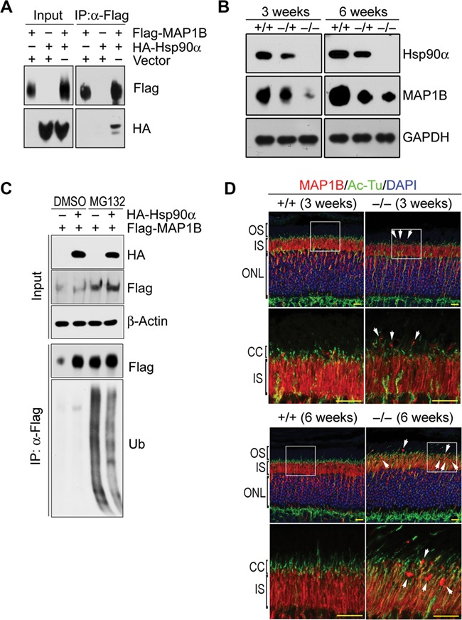
Figure 6.

MAP1B reduction in the retina of Hsp90α-deficient mice. (A) Interaction between Hsp90α and MAP1B. HA-tagged Hsp90α and Flag-tagged MAP1B were co-expressed in HEK293T cells and MAP1B was immunoprecipitated by anti-Flag affinity gel. (B) MAP1B reduction in the retina of Hsp90α-deficient mice. The retinas of wild-type (+/+), heterozygous (−/+), and homozygous (−/−) Hsp90α-deficient mice at the age of 3 or 6 weeks were isolated for western blot. (C) Decreased MAP1B ubiquitination by Hsp90α co-expression. Flag-tagged MAP1B was expressed in HEK293T cells with or without the co-expression of HA-tagged Hsp90α. MAP1B ubiquitination was analyzed after cells were treated with proteasome inhibitor MG132. Flag-tagged MAP1B was immunoprecipitated by anti-Flag affinity gel (Flag) and blotted by anti-ubiquitin antibody (Ub). (D) Immunofluorescence staining for MAP1B on retinal sections of wild-type (+/+) or Hsp90α-deficient mice (−/−) at the age of 3 or 6 weeks, respectively. The boxed areas are enlarged to show details. Arrows indicate MAP1B clusters. Scale bar, 10 μm.
