Figure 2.
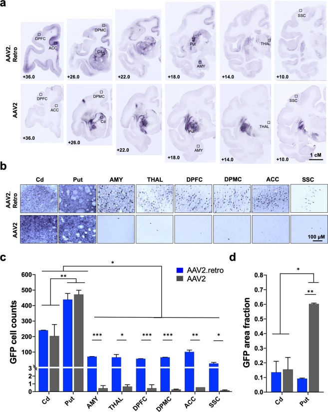
AAV2.retro-mediated retrograde transport is significantly higher compared to the parent serotype, AAV2. (a) Low and (b) high power photomicrographs of eGFP-stained, rhesus macaque coronal brain sections following injection of AAV2.retro-eGFP (top panel) or AAV2-eGFP (bottom panel) into the caudate and putamen. Rectangles in (a) illustrate the brain regions selected for high power photomicrographs displayed in (b). (c) Cell count analysis demonstrating significantly more GFP+ cells in the putamen compared to the caudate, irrespective of serotype, as well as significantly more GFP+ cells detected in extra-striatal regions in animals injected with AAV2.retro-eGFP compared to AAV2-eGFP. (d) Area fraction fractionator analysis showing a significantly higher area fraction of GFP positivity in the putamen in animals injected with AAV2-eGFP compared to AAV2.retro-eGFP. Error bars in both graphs represent standard error of the mean (SEM). Abbreviations: ACC (anterior cingulate cortex), AMY (amygdala), Cd (caudate), DPFC (dorsal prefrontal cortex), DPMC (dorsal premotor cortex), Put (putamen), SSC (somatosensory cortex), THAL (thalamus). *p < 0.05, **p < 0.01, ***p < 0.001. Scale bar 2a = 1 centimeter, Scale bar 2b = 100 microns. Graphs were made in GraphPad Prism for Mac, Version 8.

