Figure 4.
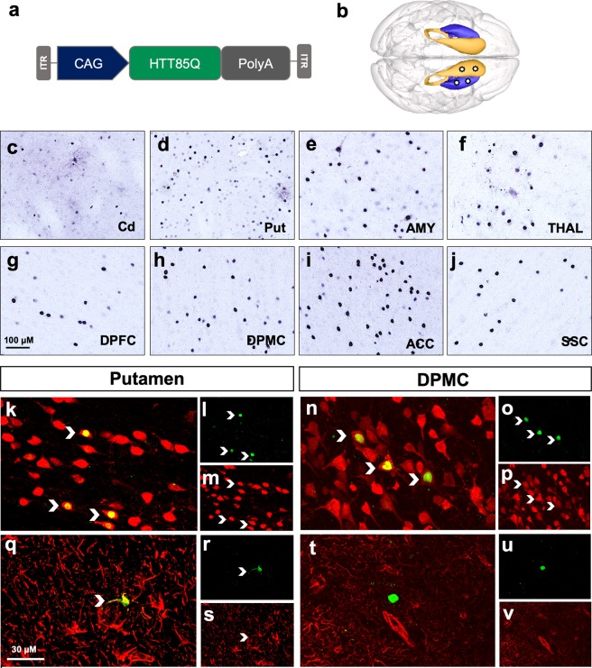
AAV2.retro-mediated delivery of mHTT into the rhesus macaque striatum leads to aggregate formation in many disease-relevant brain regions. (a) AAV2.retro vector cartoon and (b) surgical injection graphic depicting unilateral injection sites of AAV2.retro-HTT85Q into the caudate and putamen. 1-82aa staining of mHTT protein showing mHTT+ aggregates in the caudate (c), putamen (d) and several cortical and subcortical brain regions. Examples shown here include the AMY (e), THAL (f), DPFC (g), DPMC (h), ACC (i) and SSC (j). Double label immunofluorescence of HTT 1-82aa/NeuN (k–p) and HTT 1-82aa/GFAP (q–v) in the injection site (putamen, left panel) and a distal brain region (DMPC, right panel). Transduced neurons and astrocytes are indicated with chevrons. Abbreviations: ACC (anterior cingulate cortex), AMY (amygdala), Cd (caudate), DPFC (dorsal prefrontal cortex), DPMC (dorsal premotor cortex), Put (putamen), SSC (somatosensory cortex), THAL (thalamus). Scale bar in g = 100 microns, scale bar in q = 30 microns. Brain graphic in 4b was made using the 3d Brain Composer feature on the Scalable Brain Atlas website (https://scalablebrainatlas.incf.org/composer/?template=CBCetal15).

