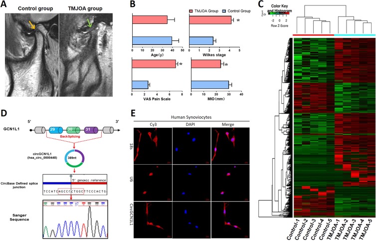
Fig. 1. CircGCN1L1 expression and localization in TMJ synovial tissues and cells.
a Representative MRI images of TMJOA and control patients. b Age, Wilkes Stage, MIO, and VAS pain score of TMJOA and control patients. N = 40 (20 different patients in each group). *p < 0.05. c Heat map of all differentially expressed circRNAs between TMJOA and control synovial samples. N = 10 (five different samples in each group). d Comparation of circGCN1L1 sequences acquired from circBase and UCSC Genome Browser with the GCN1L1 mRNA sequences. e Images of RNA FISH in synoviocytes from control patients. CircGCN1L1 probes were labeled with Cy3. Nuclei were stained with DAPI. Scale bar, 20 μm. U6 was used as an internal control for nuclear RNA, whereas 18s served as the control for cytoplasmic RNA. N = 3 (three different samples). Data are presented as mean ± S.D. Two-tailed t-test (b) was performed. MRI magnetic resonance imaging, TMJOA temporomandibular joint osteoarthritis, MIO maximum mouth incisor opening, VAS visual analog scale, FISH fluorescence in situ hybridization, DAPI dihydrochloride.

