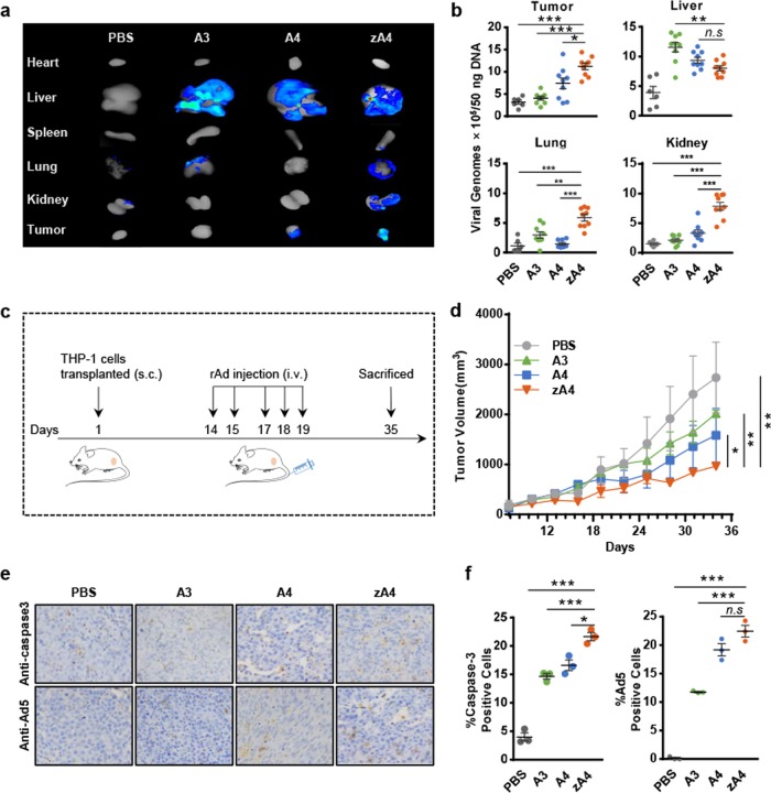
Fig. 5.
Targeting ability and antitumor effects of recombinant Ad vectors in the subcutaneous transplantation tumor-bearing model. a Biphotonic imaging of RFP in different tissues of tumor-bearing mice. Athymic nude mice were subcutaneously injected with THP-1 cells in the right flank. After the tumor volume reached ~100 mm3, the mice were i.v. injected with 5 × 10¹⁰ IU of A3, A4, or zA4. The mice were killed at 72 h after injection of the recombinant Ad and the RFP expression levels in tumor tissues and organs were analyzed by bioluminescent imaging. b The recombinant Ad in the tumor, liver, lung, and kidney was quantified by quantitative PCR. c Treatment scheme of the xenograft tumor model. d Tumor growth curve of the subcutaneous tumor-bearing mouse model. Tumor volumes were measured every 3 days and the calculated volumes (mean ± SEM) are presented in the growth curves. e Detection of caspase-3 activation and recombinant Ad infection in tumor tissues. Mice infected with recombinant Ad were killed at day 35 following tumor transplantation. The activation of caspase-3 and infection with recombinant Ad were detected by immunohistochemistry. f Statistical analysis of immunohistochemistry results. The percentages of cells positive for active caspase-3 fragments and Ad5 infection were determined in three microscopic fields in three different mice at ×200 magnification (mean ± SEM). Error bars represent the SEM. *P < 0.05; **P < 0.01; ***P < 0.001

