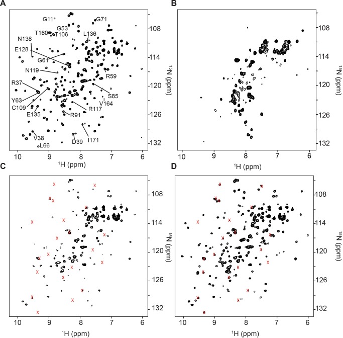
Fig 1. Protein quinary interactions are lost when the ribosome is destabilized.
A) 1H-15N HSQC NMR spectrum of 10 μM purified [U- 15N] γD-crystallin in NMR buffer. B) [U- 15N] γD-crystallin overexpressed in E. coli cells. Note the extensive loss of signals. Most of the peaks are from 15N labeled metabolites. 1H-15N HSQC NMR spectra of 10 μM purified [U- 15N] γD-crystallin in C) E. coli cell lysate containing 10 mM EDTA. Peaks that broadened in the lysate are indicated by x; and D) E. coli lysate containing 10 mM EDTA treated with 1 mM RNase A for 1 h. The majority of previously broadened peaks, x, are recovered. All spectra are shown at the same contour level.

