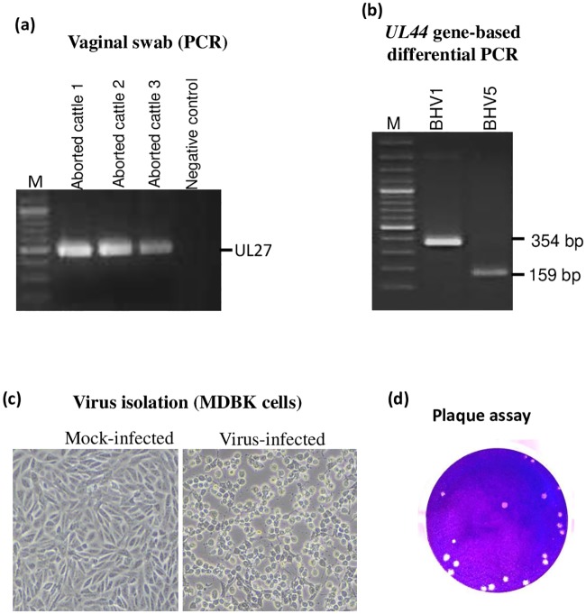
Fig 1. Isolation and identification of the agent.
(a) Amplification of BoHV1 genome in vaginal swab. Virus was recovered from the vaginal swabs in DMEM followed by DNA extraction and PCR to amplify UL27 gene of BoHV1. (b) BoHV1/BoHV5 differential PCR. PCR was carried out to amplify UL44 gene as per the method described by Claus et al. PCR amplification of UL44 from BoHV1 (reference) and BoHV5 (sample) resulted in amplification of 354 bp and 159 bp fragments respectively. (c). Virus isolation in MDBK cells. Virus recovered from the vaginal swab was used to infect MDBK cells. Cytopathic effect was observed at 3rd blind passage. Virus-infected and mock-infected cells are shown. (d) Plaque assay. Confluent monolayers of MDBK cells were infected with 10-fold serial dilutions of the virus for 1 h at 37°C followed by replacing the medium with an agar-overlay. The plaques were visible at 5–6 dpi. The agar-overlay was removed, and the plaques were stained by 1% crystal violet. The viral titers were measured as pfu/ml.

