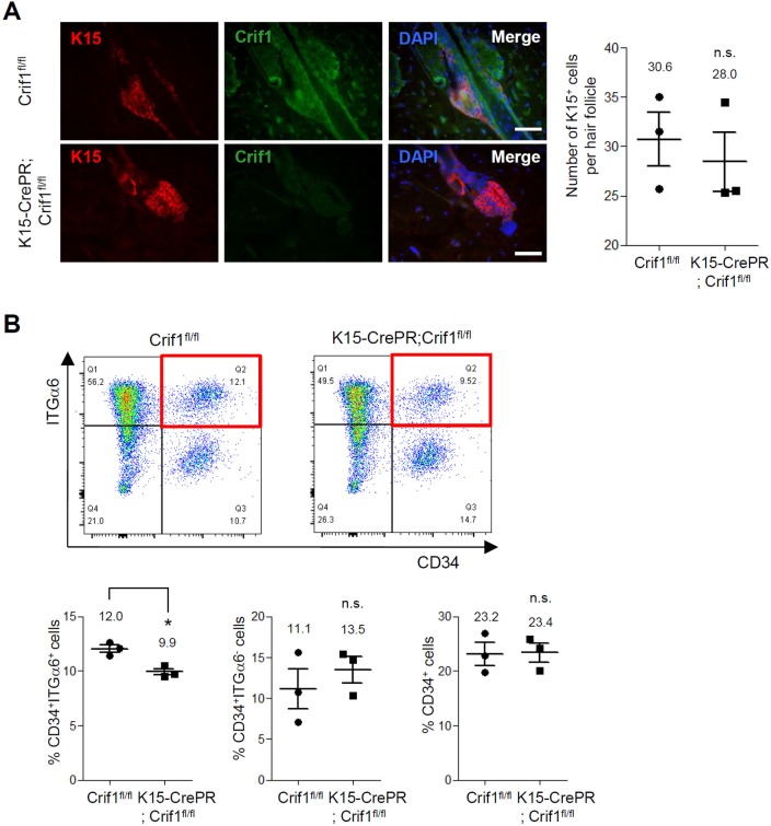
Fig 4. Effect of Crif1 deficiency on maintenance of hair follicle stem cells.
(A) Crif1fl/fl (WT) mice and K15-CrePR;Crif1fl/fl (Crif1 K15icKO) mice were shaved at P21 and then topically treated with RU486 (1 mg/mice) for 5 days. Skin sections were prepared at P44 and double-stained with K15 (hair follicle stem cell marker) and Crif1 antibodies (red, K15; green, Crif1; blue, DAPI). Scale bar, 200 μm. The number of K15-positive cells per hair follicle was quantified (n = 3, n.s.: not significant). (B) Representative flow cytometry dot plots of epidermal cells labeled with CD34 and integrin-α6 antibodies. The CD34+/integrin-α6+ cells in WT and Crif1 K15icKO mice were highlighted in red box. Percentages of CD34+/integrin-α6+ cells (lower left), CD34+/integrin-α6- cells (lower middle), and CD34+ cells (lower right) are represented in bar graph (n = 3, *P < 0.05).

