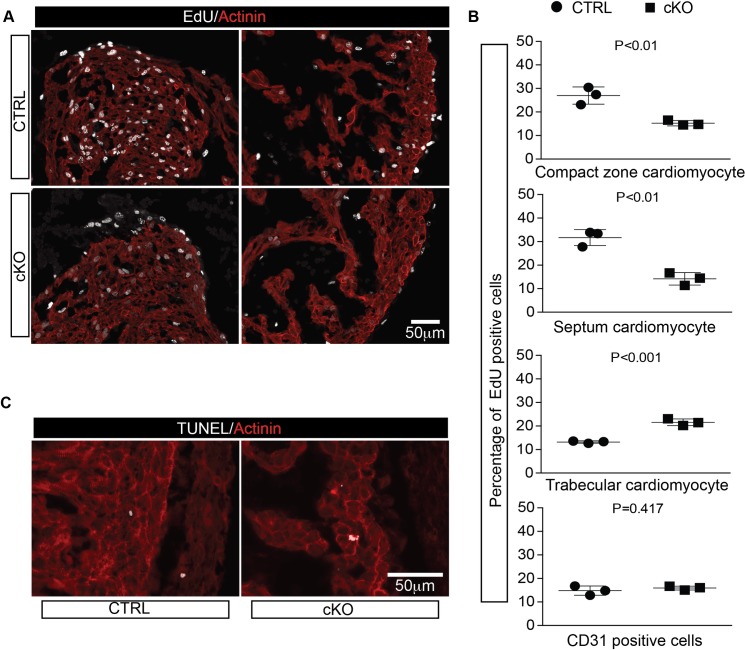
Fig 5. Loss of OGT alters cardiomyocytes proliferation.
A, EdU assay analysis of ctrl and cKO hearts at E12.5. Staining for EdU (gray) and alpha-actinin (red) are shown. B, Quantitative analysis of EdU positive rate for compact zone cardiomyocytes, septum cardiomyocytes, trabecular cardiomyocytes, and CD31 positive endothelium cells. N = 3 for each group. C, Representative images of TUNEL staining of transverse sections of ctrl and cKO hearts at E12.5. Staining for TUNEL (gray) and alpha-actinin(red) are shown.

