
Figure 1. Detection of HHV-6 in MSBB and ROSMAP RNAseq datasets.
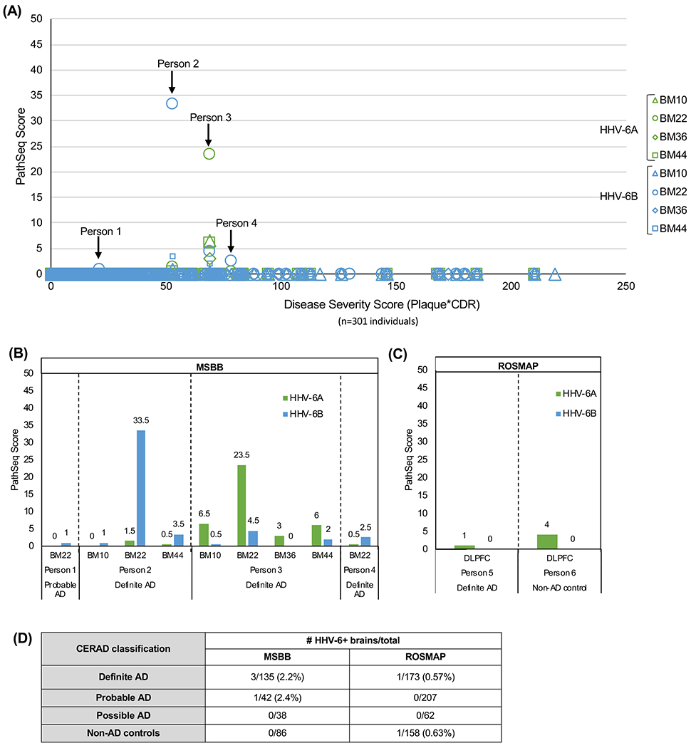
(A) HHV-6A and -6B PathSeq scores are displayed in order of increasing disease severity score (calculated as neuritic plaque density*Clinical Dementia Rating score). (B) HHV-6 was detected in 9 specimens from 4 individuals in the MSBB cohort, with varying PathSeq scores. (C) HHV-6A was detected in 2 individuals in the ROSMAP cohort, both with extremely low PathSeq scores, while no HHV-6B was detected. (D) Brains with HHV-6 in one or more brain regions are classified by CERAD neuropathology, with HHV-6 detected in individuals with definite and probable AD in addition to 1 non-AD control brain.
Key: BM10= Brodmann area 10, anterior prefrontal cortex; BM22= Brodmann area 22, superior temporal gyrus; BM36=Brodmann area 36, parahippocampal gyrus; BM44= Brodmann area 44, inferior frontal gyrus. DLPFC=dorsolateral prefrontal cortex