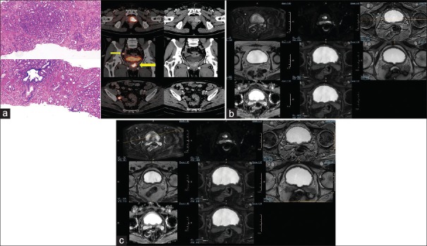
Figure 1.
(a) A 68-year-old male, serum prostate-specific antigen 76.8 ng/ml, transrectal ultrasound-guided biopsy: adenocarcinoma with Gleason score-4 + 3. Ga-68 prostate-specific membrane antigen-11 positron-emission tomography/computed tomography image shows an enhancing lesion noted in the left lobe of the prostate gland involving the posterior and anterior peripheral zones in the apical, mid glandular, and basal regions and the periurethral central zone with extension into the right lobe. There is evidence of contiguous extension of the lesion into the left seminal vesicle (thin arrow) with Ga-68 prostate-specific membrane antigen-avid metastatic right internal iliac lymph node (thick arrow). (b) Corresponding axial multiparametric magnetic resonance imaging (T2-weighted, T1-weighted, 3D VISTA SPIR, BTFE, DWI, and m-Dixon) images could not categorically delineate the left seminal vesicle. (c) Corresponding axial multiparametric magnetic resonance imaging (T2-weighted, T1-weighted, 3D VISTA SPIR, BTFE, DWI, and m-Dixon) images could not categorically delineate the right internal iliac lymph node

