
Fig. 4. NALCN voltage sensitivity primarily arises from S4 charges in domains I and II.
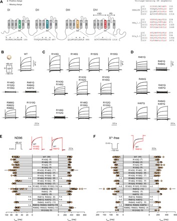
(A) Left: Schematic illustration of the structural topology of NALCN, which consists of four domains (DI to DIV) connected via intracellular linkers. Each domain contains six transmembrane segments (S1 to S6), with S1 to S4 forming the VSDs and S5 to S6 forming the pore domains. The S4 segments typically carry several positively charged residues important in voltage sensing. The conserved and missing (compared to canonical VSDs) positively charged residues in each S4 segment of NALCN are highlighted. Right: Alignment of the S4 segments of the four homologous domains in hNALCN, hNaV1.1, and hCaV2.1. Positively charged side chains are highlighted in red. (B) Current traces and Iss-V plots from X. laevis oocytes expressing WT and charge-neutralized mutants in S4 of DI to DIV using the indicated protocol. (C and D) Representative traces of single and double charge-neutralized mutants in S4 of VSDI (C) and VSDII (D). (E and F) Slow and fast time constants of depolarization-elicited currents (0 to +80 mV) in ND96 (E) and hyperpolarization-elicited currents (0 to −100 mV) in X2+-free buffer (F) for WT and VSD mutants. Superimposed traces of WT (black) and selected VSD mutants (red) are shown above the bar graphs. Data are shown as mean ± SD; *P < 0.05; **P < 0.01; ****P < 0.0001; one-way ANOVA, Dunnett’s test (against WT); n.e., no effect; n.d., not determined; gray dashed lines indicate 0 nA; numbers in parentheses indicate number of individual cells used for recordings.