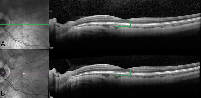
Fig. 1.

Spectral domain optical coherence tomography at first presentation (A) and follow-up visit (B). Left eye. Ellipsoid and interdigitation zone were not well-defined at the first visit; however, they improved after vitamin A treatment (green square in A and B).
