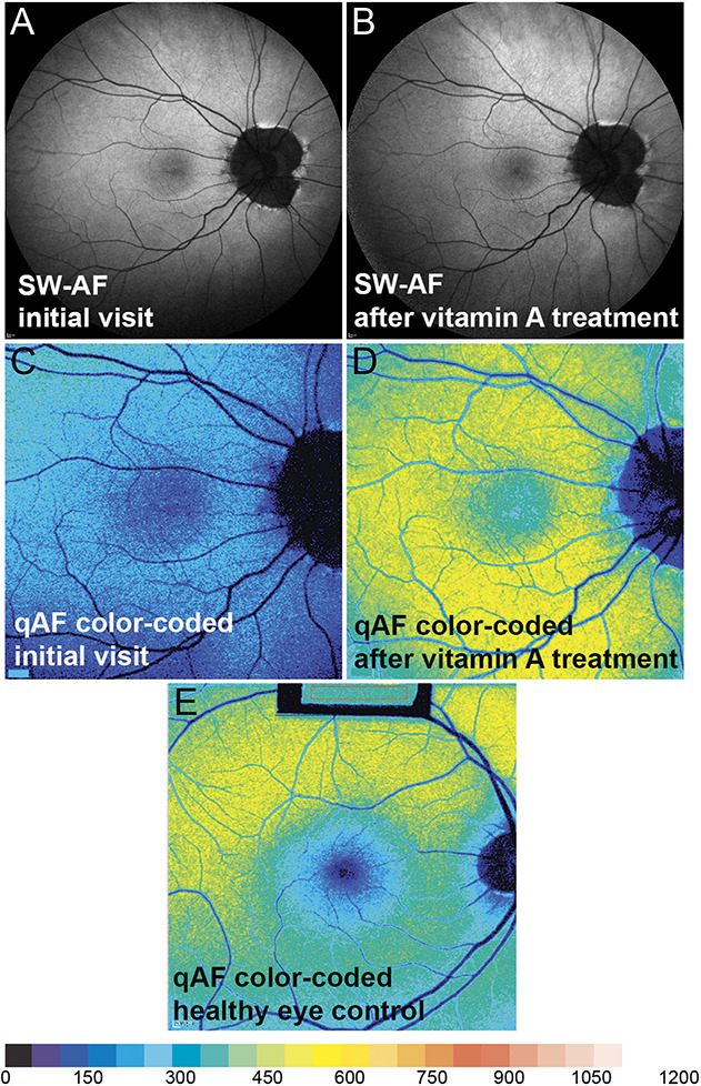
Fig. 2.

Short wavelength fundus autofluorescence and color-coded qAF. Short wavelength fundus autofluorescence (488 nm; normalized, right eye) image acquired at the initial visit (A) and after vitamin A treatment (B). Quantitative fundus autofluorescence color-coded image before (C) and after (D) vitamin A treatment. Quantitative fundus autofluorescence color-coded image of healthy control eye, 55 year-old subject (E). Note that macular pigment (blue central zone) is appreciably reduced initially and at follow-up (C and D).
